
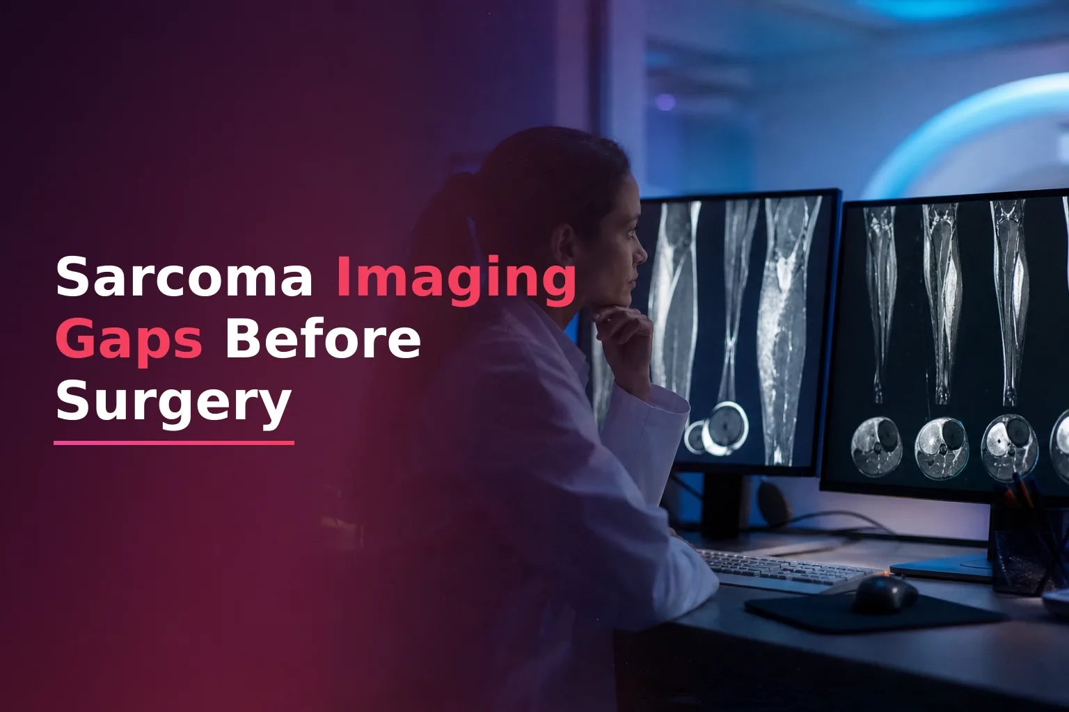
The Gap Nobody Scheduled
In soft tissue sarcoma care, the handoff from neoadjuvant chemotherapy to surgery is one of the most important transitions in oncology. The surgeon needs to know whether the tumor has responded. The imaging needs to show the tumor's current state, not its state three cycles ago. And the surgical team needs that information early enough to adjust the resection plan.
In practice, this handoff often fails. The pre-surgical imaging is ordered late, arrives the day before surgery, or ends up in a system the surgeon can't access. Surgeons sometimes enter the operating room without an up-to-date MRI showing the post-chemotherapy tumor size and tissue edges.
This article looks at why the gap happens, what research says about imaging at this point, and how oncology platforms can fix the coordination problem.
Why the Pre-Surgical MRI Is Not Optional
Soft tissue sarcomas don't always respond to neoadjuvant chemotherapy in ways that show up on physical exams or simple measurements. Tumors can develop internal necrosis, fibrosis, or cystic changes that change their biology without changing their size. Standard size-based criteria (RECIST) can therefore give the wrong picture of whether a tumor actually responded.
The 2024 guidelines from the European Society of Musculoskeletal Radiology (ESSR) are clear: imaging right after neoadjuvant therapy in soft tissue sarcoma is essential to determine how the tumor responded and to plan the surgery. MRI is the best way to assess the local area. The guidelines also say that follow-up scan timing should depend on the tumor grade and type. Pre-surgical scans are not routine studies that can wait in a scheduling backlog.
Recent research adds more details. A study found that measuring necrosis (dead tissue) on MRI after chemotherapy helps predict how the tumor will respond in soft tissue sarcoma, and this matches what doctors find during surgery. Another approach using detailed MRI analysis has shown it can predict how the tumor responds before surgery by tracking imaging changes across treatment cycles. But these findings only matter if the imaging is done before the surgery date.
The Mechanics of the Gap
The time between the last chemotherapy cycle and planned surgery must allow for bone marrow recovery, lower infection risk, and coordination between medical oncology and surgery teams. It also needs to include time for the pre-surgical scan. When these tasks aren't linked in a shared system, the imaging is most likely to get delayed.
Here is how the gap typically emerges in real clinic workflows:
- The medical oncologist finishes the final chemotherapy cycle and sends the patient to surgery. The imaging order may be understood from the referral but isn't formally ordered with a surgery-planning label or a due date.
- Radiology scheduling works from a separate system. Without a priority flag tied to the surgery date, the MRI gets scheduled at normal wait times instead of urgent pre-surgery slots.
- When the scan finishes, the report goes to whoever ordered it, usually the medical oncologist, not the surgeon. The surgical team doesn't get a notification.
- The surgical team may proceed based on the most recent available scan, which might be from partway through treatment instead of after treatment.
This is not a lack of knowledge. Every clinician understands pre-surgical imaging matters. The problem is structural: the imaging task has no clear owner, no deadline tied to the operating room, and teams can't see what's happening.
Research on oncology EHR workflows supports this. A study found that workflow differences across care settings directly affect data quality in cancer records, and that data in radiology and pathology reports aren't shared consistently across teams. When the imaging system is separate from the surgical schedule system, the handoff depends on manual communication.
What Staging at This Interval Should Include
ESSR 2024 guidance says that imaging after chemotherapy in soft tissue sarcoma should include an MRI of the main tumor to measure how much is left, look for tissue changes from treatment, and see how close it is to blood vessels and nerves. For high-grade tumors, a chest CT remains the standard way to look for lung cancer spread.
Advanced MRI protocols, which include special imaging of water movement and contrast flow, add information beyond just tumor size. These detailed measurements help surgeons understand how much living cancer remains. This matters because surgery margins are planned based on how large the cancer looks, and what MRI shows about cancer activity is at least as important as simple size measurements.
The practical point is that a pre-surgery imaging checklist for soft tissue sarcoma patients who finished chemotherapy should include at least: an advanced MRI of the main tumor, a chest CT if recent scan isn't available, and a radiology report comparing the new scan to the starting scan. Clinics without a formal checklist are most likely to miss the imaging.
The Coordination Problem Is a Scheduling Problem
Most oncology scheduling systems treat the chemotherapy-to-surgery handoff as two separate steps: a medical oncology step ending when chemotherapy finishes, and a surgery step beginning on the surgery date. There is no shared system that connects the imaging to both dates.
Consider what a coordinated workflow actually requires:
- When the final chemotherapy cycle is recorded, the system should automatically create an imaging task with a due date based on the surgery date.
- The imaging order should have a flag that both radiology and surgery can see, not just the doctor who ordered it.
- When the MRI report is finished, both the medical oncologist and the surgeon should get a notification at the same time.
- If the imaging isn't done by a certain date before surgery, the clinic operations person should see a flag on a dashboard, not find out during pre-surgery planning.
This is the kind of shared visibility across teams that modern oncology platforms can provide. The same principles apply to other treatment transitions, as shown in imaging surveillance gaps for metastatic urothelial cancer and esophageal chemoradiation toxicity monitoring.
How Rucja Addresses This Transition
Rucja's treatment timeline view shows each patient's care path as an ordered sequence of steps, not separate appointments. When a medical oncologist records the end of a chemotherapy protocol, the platform starts a pre-surgery checklist with the imaging order, a due date for radiology, and notifications to the surgery team.
When the MRI report arrives, Rucja's Lab Intelligence module pulls out key data from the radiology report and shows the main findings - residual tumor size, signal change from the baseline scan, and notes on margin distance - as a summary in the surgery planning view. The surgeon can review this in about 30 seconds instead of searching through a PDF in a separate system. To learn more about how AI extracts data from oncology reports, see how AI lab extraction changes clinical routine.
The system records when the imaging was ordered, completed, reviewed, and by whom. If surgery is within five days and the imaging hasn't been marked reviewed, the case appears in the clinic's to-do list. This is workflow accountability, not clinical decision support. In soft tissue sarcoma care, that accountability often means the surgeon has an up-to-date scan instead of an old one in the operating room.
Histologic Response Remains the True Endpoint
Pre-surgical imaging shapes the resection plan but doesn't replace the pathologist's assessment of how the tumor responded during surgery. The amount of living cancer remaining after treatment is one of the most reliable signs of how the patient will do, and it's only known after the specimen is examined.
Pre-surgical imaging helps the surgical team know what to expect. A tumor that appears to have responded may allow different margins than one that appears stable or enlarged. Either way, the imaging must be up to date, complete, and available to the surgery team before the patient enters the operating room.
You can schedule a 30-minute demo to see the neoadjuvant-to-surgery workflow with your hospital's data, including the imaging task checklist and cross-team notifications. Book a demo to learn how Rucja manages your sarcoma care.